
What is the difference between GC – MS Full Scan and Selected Ion Monitoring?
In order to understand the difference between mass spectroscopic full scan and selected ion monitoring it is essential for you to have a clear concept of spectral scanning in optical spectroscopy as both have several commonalities.
Scanning in optical spectroscopy
Optical spectroscopy serves as a valuable tool for qualitative and quantitative estimation of constituents of light absorbing samples. The sample is taken in a fixed path length cell and irradiated with light covering the wavelengths of interest. Generally in the UV- Visible region a deuterium lamp serves as a light source for the UV region and a tungsten- halogen lamp for the visible region. A monochromator using a grating isolates the required wavelengths which irradiate the sample at a rate dependent on the scan rate selected.
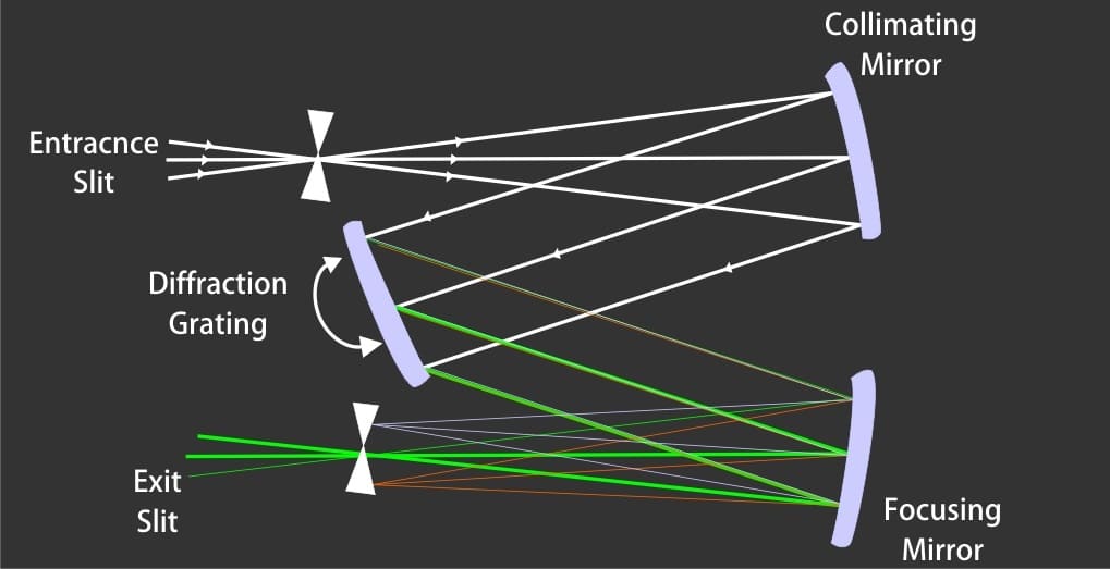
Schematic diagram of a monochromator
The full scan over the selected wavelength range provides several absorption peaks reflecting presence of light absorbing species in the sample. In order to quantitate any particular species the monochromator is set at a particular wavelength and the absorbance intensity recorded to arrive at the concentration using the Beer Lambert law of light absorption.
Scanning in mass spectrometry
In GC – MS analysis the mixture components separated by the GC column reach the ionization chamber of the mass spectrometer. The analytes are subjected to high energy electron beam which results in ionization and fragmentation of parent molecules. The resulting ionized species are led into the quadrupole mass analyzer which can be compared to the monochromator in the optical spectrometer. The monochromator isolates specific wavelengths reaching it from the broadband light source whereas the quadrupole resolves the ionic species on the basis of their mass to charge ratios (m/z).
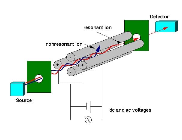
Quadrupole mass filter schematic diagram
Mass separation is accomplished by application of a DC voltage and RF frequency at diagonally opposite ends of the quadrupole rods. The RF frequency is scanned over a range to permit specific m/z value fragments to pass across the quadrupole rods and focus on the mass sensitive detector.
Full scan monitoring
Full scan monitoring provides a qualitative picture of the composition of the sample. It involves scanning the mass range beginning at the smallest mass of fragment ions to the highest mass expected for the fragments. Typically the mass range selected would be from, say 50 to about 400, depending on what you would anticipate in your sample taking care to eliminate possible interferences from sample matrix and atmospheric gases. Too low mass settings could result in interference from nitrogen (m/z = 28) or carbon dioxide (m/z=44) present in the air. In case a very large mass range is selected the sensitivity would be lower as fewer scans per second would be performed. At the same time information on masses expected outside the expected mass fragments range would be of little use to you. In a nutshell it is necessary to run a full scan of your sample before you limit your search for identification and quantification of species of interest in the sample.
Selected ion monitoring
The full scan mode is useful for qualitative detection, mass scouting and for studies on the fragmentation patterns of unknowns. On the other hand, selected ion monitoring helps in quantitative studies on the specific ions of interest in the spectra of compound (s) of your interest.
In the selected ion monitoring mode data is collected on only the selected masses of interest rather than over wide range of masses. This improves the specificity of the instrument resulting in 10 -100 fold increase in detection limits. This feature helps eliminate interferences arising due to complex sample matrices thereby contributing to the potential of mass spectroscopy as an analytical technique.
What is the difference between full scan and selected ion monitoring in GC-MS?
Full scan mode records data across a wide mass range and is useful for qualitative detection, mass scouting, and studying fragmentation patterns of unknowns. Selected ion monitoring instead collects data only on selected masses of interest, which improves specificity and is used for quantitative studies.
When should I use full scan mode in GC-MS analysis?
It is necessary to run a full scan of the sample before limiting the search to specific masses, since full scan provides a qualitative picture of the sample composition and reveals fragmentation patterns of unknown compounds.
Why does selected ion monitoring improve detection limits?
By collecting data only on selected masses rather than scanning a wide range, selected ion monitoring improves instrument specificity, resulting in a 10 to 100 fold increase in detection limits and helping eliminate interferences arising from complex sample matrices.
What mass range is typically selected for a full scan?
The mass range selected is typically around 50 to 400, depending on what is anticipated in the sample, chosen to avoid interference from atmospheric gases such as nitrogen at m/z 28 or carbon dioxide at m/z 44.
How does the quadrupole mass analyzer resolve ions in GC-MS?
Mass separation is accomplished by applying a DC voltage and RF frequency at diagonally opposite ends of the quadrupole rods, with the RF frequency scanned over a range to let specific m/z fragments pass through and reach the detector. This role is comparable to how a monochromator isolates specific wavelengths in optical spectroscopy.
Why is selecting too large a mass range for a full scan not ideal?
Selecting a very large mass range lowers sensitivity because fewer scans per second are performed, and masses outside the range actually expected in the sample provide little useful information anyway.